Part 2: Proactive steps for breast cancer risk reduction: What type of breast screening should I get?
In this article we will talk about the different types of breast screening available. First, let’s try to minimize any radiation fears. If you’ve wondered if your mammogram may be bad for you because of the radiation, consider this: The amount of radiation to your breasts will be less than a “normal x-ray.” The “dose” will be about 0.4 millisieverts. You typically get that much radiation just being in your natural surroundings (indoors/outdoors) over seven weeks. For nearly all woman, the benefits of getting your mammogram outweigh this minor amount of radiation exposure.
We will discuss the various types of mammograms (yes, there are a few variations depending on availability at the clinics near you) as well as other techniques to screen that you may not have heard of before.
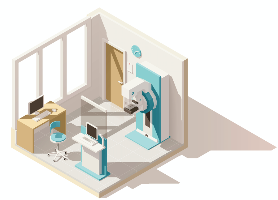
Mammograms are a technique used to take images of your breasts after squishing them between two plates. (To enjoy some mammogram humor, click here.) There are Traditional Mammograms, Digital Mammograms, 3D Mammograms, and Contrast Enhanced Mammograms. Who knew there were so many different types?!
A Traditional Mammogram creates an image using an x-ray system.
Digital Mammograms are the most common screening technique currently used in the US. It replaces the traditional x-ray film with digital images that can be viewed on a computer instead of on x-ray film. The imaging process is typically faster than traditional mammography.
3D Mammograms are also referred to as Tomosynthesis. This type of screen releases the same amount of radiation as a traditional mammogram. This technology takes multiple pictures of the breast which are then used to recreate a 3D image of the breast. This type of screening technique reduces the potential for false positive callbacks, and is more accurate at detecting breast cancers compared to traditional mammograms. This type of screen can benefit any woman, particularly those with dense breast tissue, or other risk factors. All women should consider 3D mammography. Before scheduling your mammogram, see if your insurance will cover 3D mammograms, and if they do, find a breast screening clinic that offers 3D mammography.
Contrast Enhanced Mammography (CEM) is similar to a standard mammogram except that you will receive a contrast agent injected into your arm vein. Two images are taken at each position, a “low energy” image that looks like your normal mammogram, and a “high energy” image that will pick up the contrast agent you were injected with. Tumors typically “drink-up” more of this contrast agent compared to your normal breast tissue, so it will be easier to see on the mammogram image. This type of screen may be beneficial in women with dense breast tissue. It is often used on women who are being called back because of an inconclusive standard mammography result. This imaging option is more accurate than standard mammography options and has similar accuracy to breast MRI.


Lastly, Thermography uses an infrared camera to look at blood flow and heat patterns in the breast. This is not a screening substitute for mammography; it should only be paired with your mammogram screening. On its own, thermography can miss small, early stage tumors. Changes in blood flow happen as tumors grow in size because they start to trick the breast into making new blood vessels to help supply the tumor with nutrients so it can continue to grow. Relying on thermography alone means you are willing to wait until your breast tumor is big enough to see these changes occur—remember mammography and MRI can pick up these changes at an earlier stage.
Whichever screening tool you and your doctor choose to use, make sure you are being screened and being proactive about your own health.
